

ames B. Carr, M.D.
Conservative Treatments We Recommend
Conservative Management of Spinal Dysfunction: Dr. Carr is one of the few spinal surgeons in the area with the experience and expertise to effectively coordinate and oversee comprehensive programs for conservative, non-surgical management. Conservative management is always encouraged prior to any spinal surgery and consists of the following:
Patient Specific Education: As each of our patients is unique in their presentation, each deserves education specific to their condition. Using a variety of qualified ancillary providers Dr. Carr offers the unique opportunity to receive information and education that is tailored to each individual and their needs.
Physical Therapy: Dr. Carr works extensively with local physical therapists to compile the most effective rehabilitation programs available. Dr. Carr has specific parameters that he wants achieved and works with qualified Doctors in Physical Therapy as well Exercise Physiologists and Certified Athletic Trainers (at many local physical therapy offices) to achieve the highest level of function possible for his clients.
Intervention and Diagnostic Radiology: Dr. Carr insists on working with only the most current technology when it comes to radiologic procedures as well as diagnostic testing. Working with national and internationally regarded technology allows Dr. Carr to “see” things that other surgeons may not have the ability or experience to identify. Only through the use of the most advanced real time technology is Dr. Carr able to provide a specific diagnosis individual to each patient.
Pain Management: Some patients may require some degree of pain management in working to improve their quality of life. Dr. Carr offers access to specific and controlled routes of pain management through collegial interventions with appropriate pain management experts.
Maximizing Health and Wellness: Dr. Carr believes that prevention and prophylaxis are ultimately the best medicine and as such he works closely with a variety of practitioners to achieve the goal of maximizing health and wellness for his clients. Working with the sound medically based programs available through all conservative treatment plans; Dr. Carr is assured that his patients desiring continued improved quality of life after completion of a course of conservative care can meet their goals of maximizing health and reducing the risk of future injury.
Surgical Options We Recommend
Through advanced Residency and Fellowship training combined with cutting-edge training in spinal surgery, Dr. Carr is able to offer advanced surgical technique for individuals needing spinal fusion or laminectomy. Dr. Carr works with only the finest instruments and most qualified surgical teams to achieve optimal results for his patients.
Microdiscectomy
In a microdiscectomy or a microdecompression spine surgery, a small portion of the bone over the nerve root and/or disc material from under the nerve root is removed to relieve neural impingement and provide more room for the nerve to heal. A microdiscectomy spine surgery is typically performed for lumbar herniated disc.
Microdiscectomy helps leg pain
A microdiscectomy surgery is actually more effective for treating leg pain (radiculopathy) and sciatica than for lower back pain. The impingement on the nerve root (compression) can cause substantial leg pain, and while it may take weeks or months for the nerve root to fully heal and any numbness or weakness get better, patients normally feel relief from leg pain almost immediately after a microdiscectomy surgery.
Microdiscectomy spine surgery procedure
A microdiscectomy spine surgery is performed through a small (1 inch to 1 1/2 inch) incision in the mid-line of the low back.
-
First, the back muscles (erector spinae) are lifted off the bony arch (lamina) of the spine. Since these back muscles run vertically, they can be moved out of the way rather than cut.
-
The surgeon is then able to enter the spine by removing a membrane over the nerve roots (ligamentum flavum), and uses either operating glasses (loupes) or an operating microscope to visualize the nerve root.
-
Often, a small portion of the inside facet joint is removed both to facilitate access to the nerve root and to relieve pressure over the nerve.
-
The nerve root is then gently moved to the side and the disc material is removed from under the nerve root.
Importantly, since almost all of the joints, ligaments and muscles are left intact, a microdiscectomy spine surgery does not change the mechanical structure of the patient's lower spine (lumbar spine).
When to have microdiscectomy spine surgery
In general, if a patient's leg pain due to a disc herniation is going to get better, it will do so in about six to twelve weeks. As long as the pain is tolerable and the patient can function adequately, it is usually advisable to postpone back surgery for a short period of time to see if the pain will resolve with conservative (non-surgical) treatment alone.
If the leg pain does not get better with conservative treatments, then a microdiscectomy surgery is a reasonable option to relieve pressure on the nerve root and speed the healing. Immediate spine surgery is only necessary in cases of bowel/bladder incontinence (cauda equina syndrome) or progressive neurological deficits. It may also be reasonable to consider back surgery acutely if the leg pain is severe.
Microdiscectomy spine surgery is typically recommended for patients who have experienced leg pain for at least six weeks and have not found sufficient pain relief with conservative treatment (such as oral steroids, NSAID's, and physical therapy). However, after three to six months, the results of the spine surgery are not quite as favorable, so it is not generally advisable to postpone microdiscectomy surgery for a prolonged period of time (more than three to six months).
After the microdiscectomy surgery
Usually, a microdiscectomy spine surgery procedure is performed on an outpatient basis (with no overnight stay in the hospital) or with one overnight in the hospital. Post-operatively, patients may return to a normal level of daily activity quickly.
Some spine surgeons restrict a patient from bending, lifting, or twisting for the first six weeks following surgery. However, since the patient's back is mechanically the same, it is also reasonable to return to a normal level of functioning immediately following microdiscectomy spine surgery. There have been a couple of reports in the medical literature showing that immediate mobilization (return to normal activity) does not lead to an increase in recurrent lumbar herniated disc.
Microdiscectomy spine surgery success rate
The success rate for a microdiscectomy spine surgery is approximately 90% to 95%, although 5% to 10% of patients will develop a recurrent disc herniation at some point in the future.
A recurrent disc herniation may occur directly after back surgery or many years later, although they are most common in the first three months after surgery. If the disc does herniate again, generally a revision microdiscectomy will be just as successful as the first operation. However, after a recurrence, the patient is at higher risk of further recurrences (15 to 20% chance).
For patients with multiple herniated disc recurrences, a spine fusion surgery may be recommended to prevent further recurrences. Removing the entire disc space and fusing the level is the only way to prevent recurrent disc herniations.
Recurrent herniated discs are not thought to be directly related to a patient's activity, and probably have more to do with the fact that within some disc spaces there are multiple fragments of disc that can come out at a later date. Unfortunately, through a posterior microdiscectomy spine surgery approach, only about 5 to 7% of the disc space can be removed and most of the disc space cannot be visualized. Also, the hole in the disc space where the disc herniation occurs (annulotomy) probably never closes because the disc itself does not have a blood supply. Without a blood supply, the area does not heal or scar over. There also is no way to surgically repair the annulus (outer portion of the disc space).
Following a microdiscectomy spine surgery, an exercise program of stretching, strengthening, and aerobic conditioning is recommended to help prevent recurrence of back pain or disc herniation.
Microdiscectomy surgery risks and complications
As with any form of spine surgery, there are several risks and complications that are associated with a microdiscectomy spine surgery procedure, including:
-
Dural tear (cerebrospinal fluid leak). This occurs in 1% to 2% of these surgeries, does not change the results of surgery, but post-operatively the patient may be asked to lay recumbent for one to two days to allow the leak to seal.
-
Nerve root damage
-
Bowel/bladder incontinence
-
Bleeding
-
Infection
However, the above complications for microdiscectomy spine surgery are quite rare.
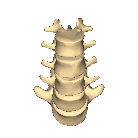
Spinal Fusion
Spinal fusion is a "welding" process by which two or more of the small bones (vertebrae) that make up the spinal column are fused together with bone grafts and internal devices such as metal rods to heal into a single solid bone. The surgery eliminates motion between vertebrae segments, which may be desirable when motion is the cause of significant pain. It also stops the progress of a spinal deformity such as scoliosis. A spinal fusion takes away some of the patient's spinal flexibility.
Most spinal fusions involve relatively small spinal segments and thus do not limit motion very much. Spinal fusion is used to treat:
-
Injuries to spinal vertebrae.
-
Protrusion and degeneration of the cushioning disk between vertebrae (sometimes called protruding disk or herniated disk).
-
Abnormal curvatures (such as scoliosis or kyphosis).
-
Weak or unstable spine caused by motion of the vertebrae, infections or tumors.
Bone is the most commonly used material to help promote fusion. Generally, small pieces of bone are placed into the space between the vertebrae to be fused. Sometimes larger solid pieces of bone are used to provide immediate structural support. Bone may come from:
-
The patient (autogenous bone).
-
A bank of bone harvested from other individuals (allograft bone).
Autogenous bone is generally considered superior at promoting fusion. But drawbacks to using it include extra surgery to remove bone from the patient's body such as the hip or pelvis. Allograft bone is available from bone banks. Other bone graft substitutes are being developed, but have yet to be proven as cost effective substitutes for autogenous bone graft for general use.
After the fusion procedure has been performed, the adjacent spinal segments are held immobile to allow fusion to progress. Immobilization is achieved through internal fixation devices or external bracing or casting. Both forms of immobilization may be necessary at times.
Risks for any surgery include bleeding and infection. Additional risks for spinal fusion surgery include urinary difficulties (retention) and temporary decreased or absent intestinal function. Patients can best prepare for spinal fusion surgery by:
-
Thoroughly consulting with their doctor before surgery.
-
Making arrangements for post-op care and assistance from family members and/or home modifications if needed.
-
Achieving good nutritional status before and after surgery.
-
Following a recommended exercise program before and after surgery.
-
Maintaining a positive mental attitude and outlook.
Dr. Carr is certified and experienced with performing spinal fusion, and has 15+ years of experience with this procedure. He performs spinal fusion at the cervical, thoracic and lumbar levels, and to correct severe scoliosis. Spinal fusion may be performed through three approaches: posterior, anterior, or transverse. Lumbar fusion may also include decompression of a nerve if nerve impingement is present due to severe degeneration of the discs. Dr. Carr’s most common fusions performed are:
-
Anterior/posterior cervical decompression and fusion
-
Lumbar decompression and fusion
-
Lumbar fusion with instrumentation
-
Posterior/ transverse/anterior lumbar interbody fusion
-
Multi-level Thoracic or Thoracolumbar spinal fusion to correct scoliosis
Artificial Disc Replacement
Dr. Carr performed the first Charite artificial disc procedure in the Central Coast of California on April 21, 2005 at Twin Cities Community Hospital. We are pleased to offer a surgical alternative to spinal fusion surgery, referred to as an artificial disc replacement. Artificial disc replacement is a procedure that involves replacing a painful disc that is causing chronic back pain with an artificial disc that provides pain relief without compromising the spine's natural anatomical structure. Artificial disc replacement surgery may be performed on the lower back (lumbar spine) or the neck (cervical spine). Artificial discs are structurally similar to the damaged discs that are replaced and share similar functions, including acting as shock absorbents in the back or neck.
Artificial discs replacement surgery is approved by the U.S. food and drug administration (FDA) for both the lumbar and cervical spine. Dr. Carr chooses to operate with the prodisc-L total disc replacement (DePuy Synthes) and Mobi-C Total Disc Replacement (LDR) models.
Artificial Disc Replacement is a surgical treatment alternative for degenerative disc disease of the cervical and lumbar spine.
Laminectomy and Laminotomy
Laminectomies and Laminotomies are surgeries that remove a small amount of bone from the spine. These procedures relieve pressure from a compressed nerve, which can greatly
During a Laminectomy, the entire lamina is removed from the affected vertebra. The opening created may be enough to remove pressure from the nerve, and if needed, bone spurs or disc matter may be removed as well to relieve pressure. After laminectomy, the opening in the spine is protected by the thick back muscles.
During a Laminotomy, a portion of the lamina is removed from the vertebra above and below the pinched nerve. The small opening created is sometimes enough to relieve pressure from the nerve, but in most cases, disc matter or bone spurs will need to be removed as well.
View this video on Youtube about Microdiscectomy Surgery courtesy of Dr. Larry Parker, M.D.:
View this video on Youtube about Fusion Surgery for Scoliosis courtesy of Bronte Hamilton:
View this video on Youtube about Cervical and Lumbar Fusion Surgery courtesy of Nucleus Medical Media:
View these videos on Youtube about Disc Replacement courtesy of Dr. Larry Parker, M.D.:
View this video on Youtube about the Mobi-C Disc Implant courtesy of LDR:
View this video on Youtube about Laminectomy courtesy of MediLaw.TV:
Coflex
The coflex® Interlaminar Stabilization® device is a single-piece titanium implant that goes in the back of your spine to treat moderate to severe spinal stenosis. It is also used above a lumbar spinal fusion to increase stability. It’s amazingly simple, very strong, and flexible enough to support your spine without having to fuse your bones together. The coflex device comes in five sizes to fit most patient anatomy, is implanted directly following a surgical decompression, and can be performed in an outpatient setting.
coflex is the FIRST AND ONLY posterior lumbar motion preservation solution with Level I evidence (the highest possible level of clinical data) from two prospective randomized studies against two treatment options (decompression alone and decompression with fusion) across two countries (U.S. and Germany). It has been implanted in more than 163,000 patients in over 60 countries worldwide.
View this video on Youtube and click on this link https://ryortho.com/2016/02/5-year-data-favors-coflex-over-fusion-for-back-pain/ to learn about Coflex Interlaminar Stabilization:
View this video on Youtube about Laminotomy courtesy of Spine Nevada:
OLIF: Oblique Lateral Interbody Fusion
A MUSCLE-SPARING, NERVE AVOIDANCE PROCEDURE
Oblique Lateral Interbody Fusion (OLIF) procedures by Medtronic offer a complete, minimally invasive solution for the correction of degenerative and deformity spinal conditions. These procedures allow access from L2 to S1 from a single position, without repositioning the patient.
By utilizing an oblique lateral approach to the spine, OLIF Procedures enable placement of a large interbody graft on to the apophysis, the strongest part of a vertebral body. The graft provides anterior column support while avoiding risks and obstacles associated with traditional anterior, posterior, and direct lateral approaches.
Watch Dr. Carr's news interview regarding the OLIF procedure here: